When you buy through link on our site , we may earn an affiliate perpetration . Here ’s how it work .
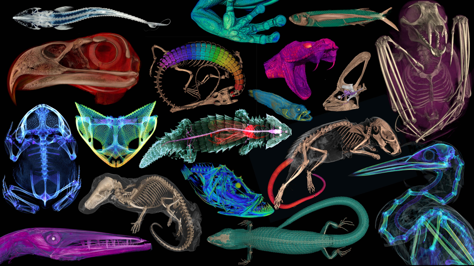
Incredible 3-D images of over 13,000 vertebrate — stand for one-half of the world ’s described genus — have been created as part of a project to make museum specimens available to all .
Fromspine - tailed mice(Acomysspecies ) torare rim rock crown snakes(Tantilla oolitica ) , natural collections from museums around the world are being tally toopenVertebrate ( oVert ) — a five - year project fund by the National Science Foundation creating a database of cypher tomography ( CT ) scans of specimens .
With CT scanning, scientists can study a specimen’s internal anatomy without the need for dissection.
CT scanscombine multiple ex - beam image taken from unlike angles around the body to create elaborated grouchy - sectional image , enabling scientist to peer through the exterior of animals without damaging the specimens . This gives them worthful brainstorm into skeletal structure .
Some specimens were even stained during skim so researchers could view soft tissue structures , revealing stomach contents , nut , parasites and electronic organ .
As of November 2023 , 13,000 specimen across 18 U.S. insane asylum have been scanned .
Using various methods, researchers can reconstruct museum specimens as digital 3D models.
Related:3D scans reveal that beetles have private pocket on their backs
The project team has released a extract of images from the project show creatures in remarkable detail . These admit a CT scan of the final meal of a hognose snake(Heterodon platirhinos ) , the prickly backbone of a four - toed Erinaceus europeaeus ( Atelerix albiventris ) and a black - bellied fruit bat ( Melonycteris melanops ) .
Whenfirst created , museum held uncommon specimens as part of individual collections of wealthy person . Now , museums are open to the populace , but some solicitation remain concealed .
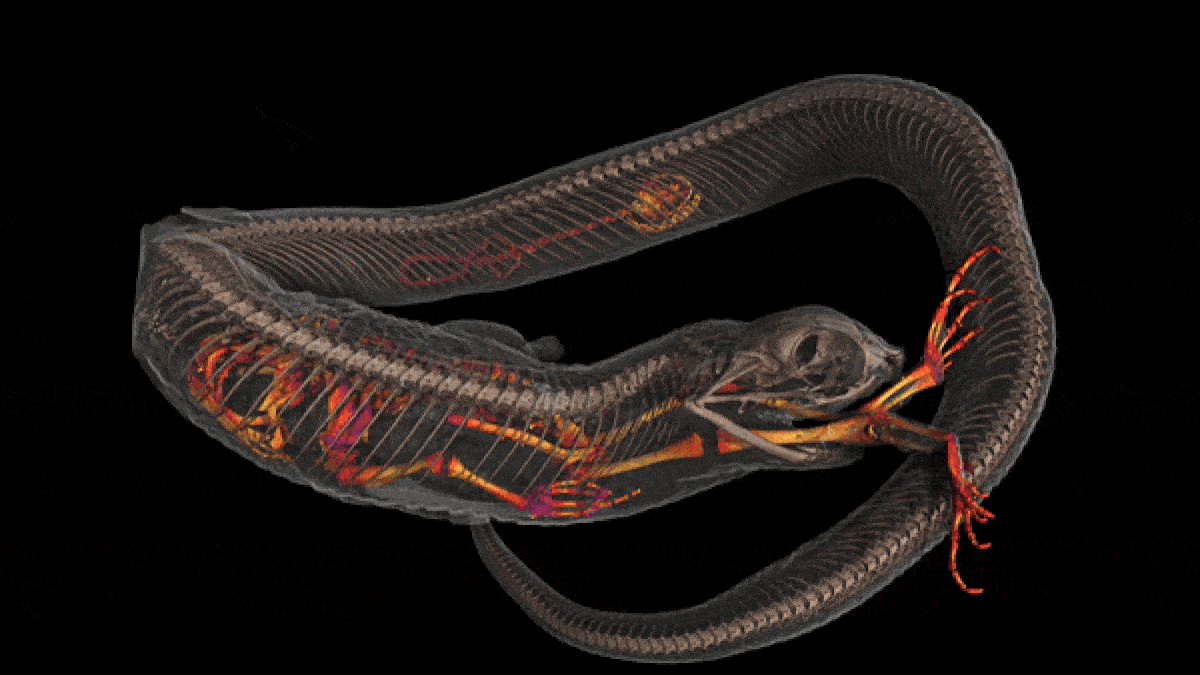
A computed tomography scan showing the final two meals of an Eastern hognose snake (Heterodon platirhinos): a salamander and a toad.
" approach to these collections is often limited ; cost of travelling , space restrictions , fragility of specimens , all can forestall people from working with these samples,“Edward L. Stanley , an writer and Director of the Digital Imaging Division at the Florida Museum of Natural History , tell Live Science in an e-mail .
— Watch a rare pink albino elephant baby performing by a waterhole in adorable footage
— Watch promising red blood - suck parasite fete on guzzler eel in rare , deep - ocean footage
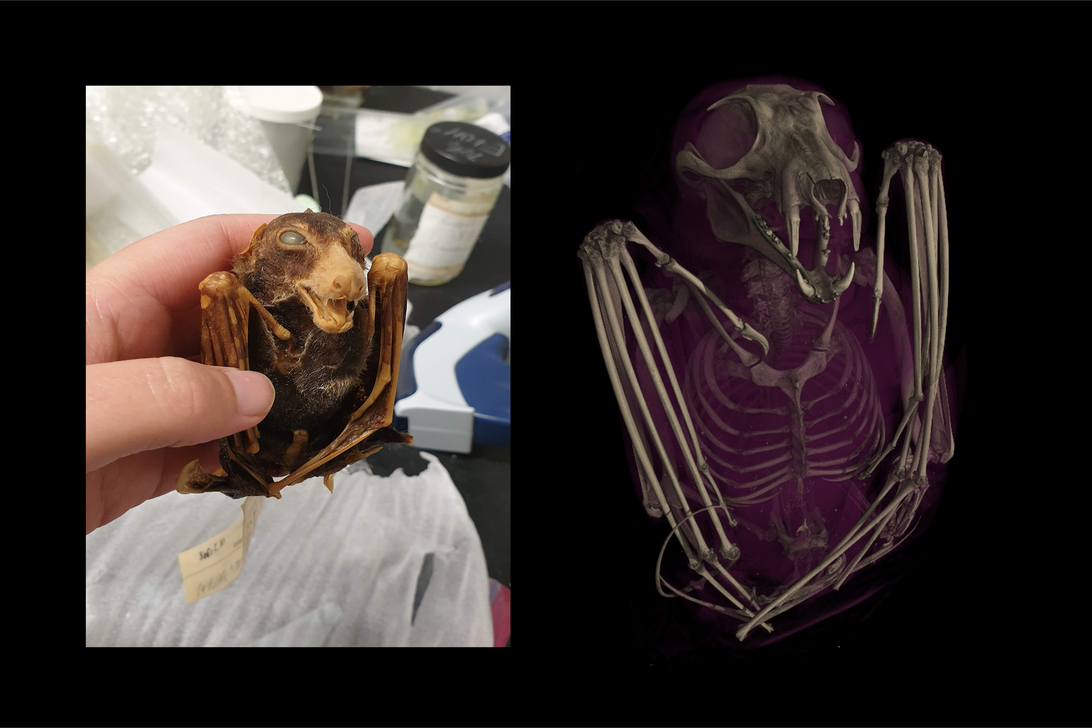
CT scan of the skeletal structure of a black-bellied fruit bat (Melonycteris melanops).
— Pangolin courtship rite and birth of a ' pangopup ' captured in incredible , rare footage
Stanley is an source of a study published Mar 6 . in the journalBioSciencethat leave a summary of the projection to engagement .
study specimens often involvesdissections and chemical substance examination , which can damage specimen , further specify handiness . " Museums have to strike a correspondence between using their specimens and using them up , which often means that rarest and most significant specimens are often the hardest to access " , Stanley said .
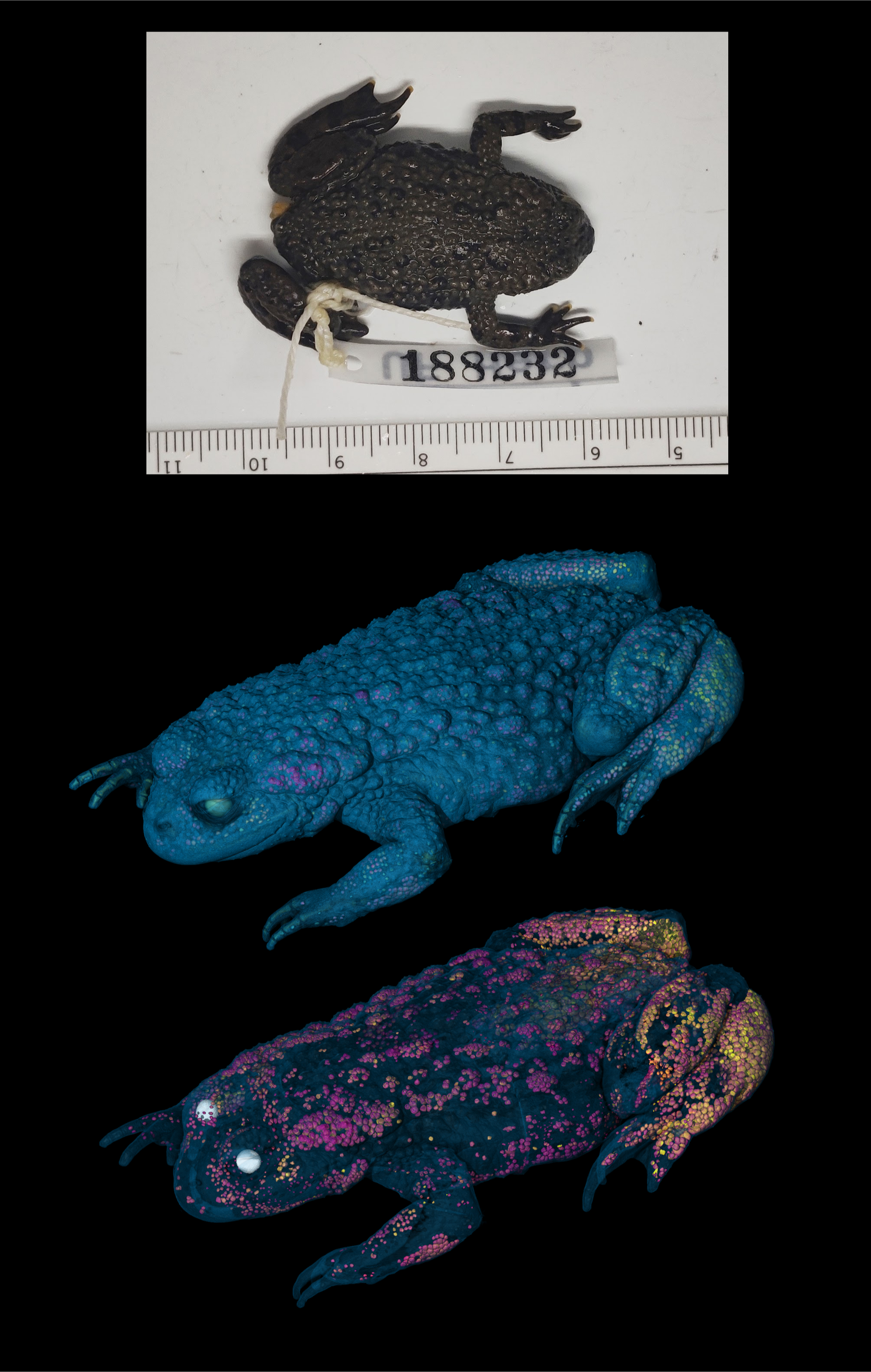
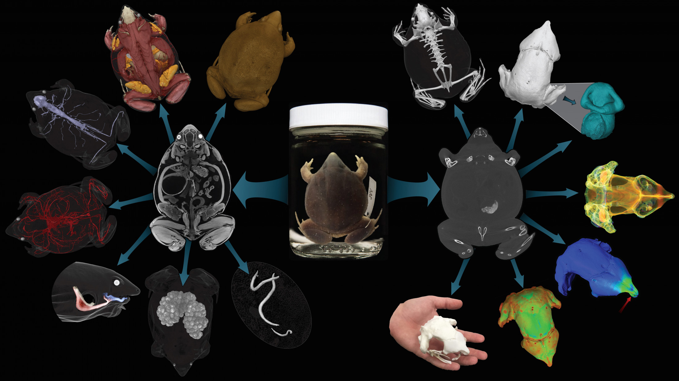
CT scan of an oriental fire-bellied toad, showing mineralization in the skin.
" The digital data point from CT scanning provides a fast and modest - risk agency for museums to make even their most delicate , rarified or irreplaceable specimen available to anyone , anywhere in the earth , " Stanley added .
image capturing a famish Panthera leo , fighting bison and perdition of vipers honor in environmental photography awards
Hoatzin : The unknown ' hoactzin ' born with clawed wings that is likely an evolutionary ' orphan '
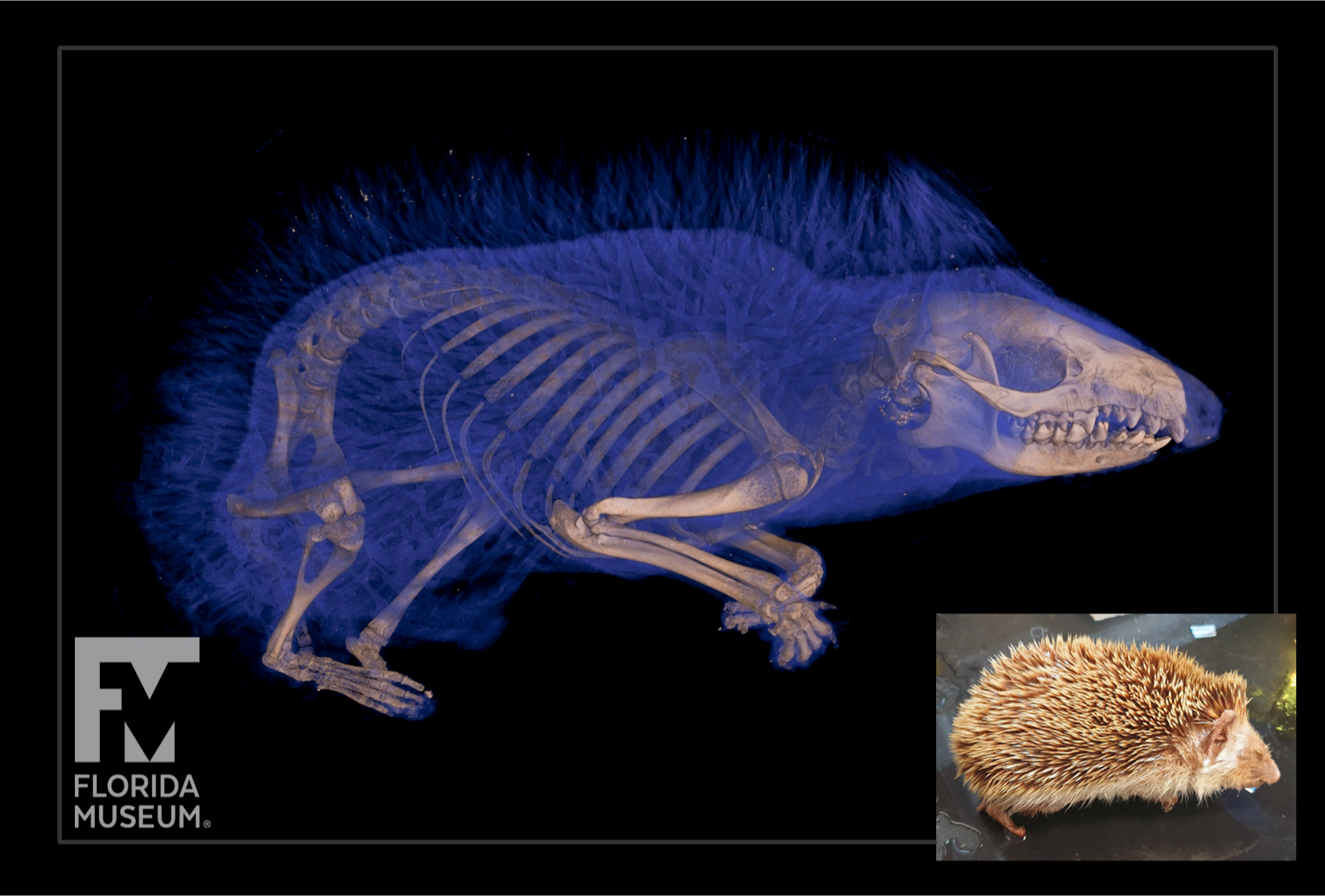
CT scan showing the skeletal structure of a four-toed hedgehog (Atelerix albiventris).
The constant surveillance of modern biography could worsen our brain function in ways we do n’t full see , stir up studies propose
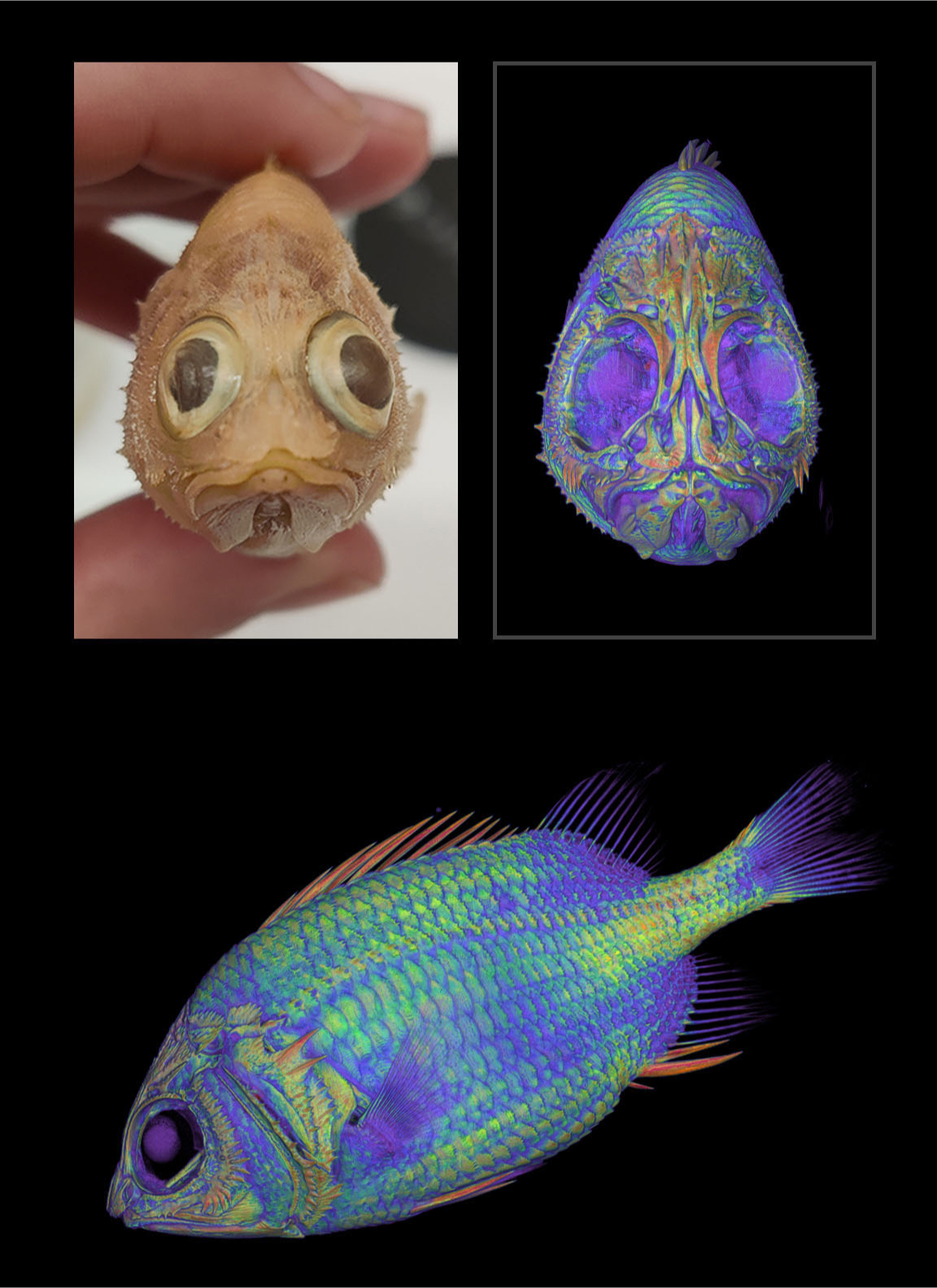
CT scan of a cardinal soldierfish (Plectrypops retrospinosus).
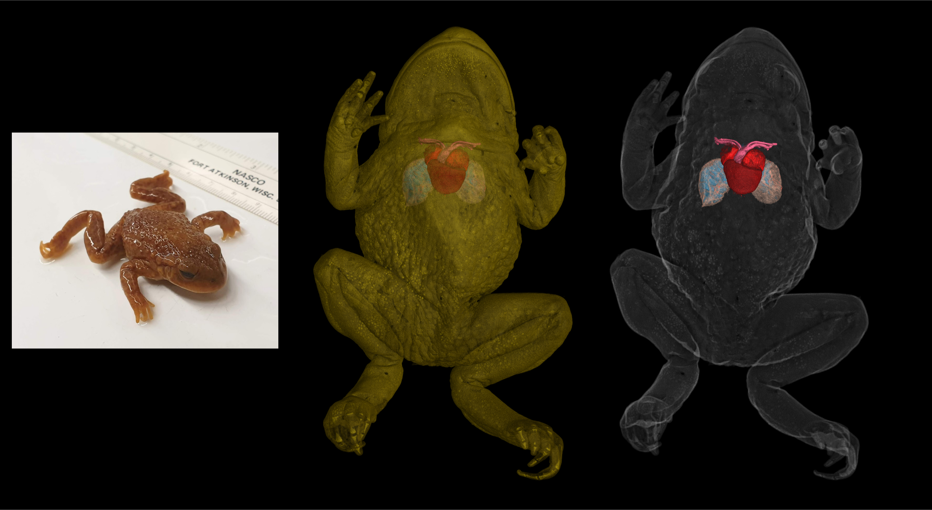
CT scan of a species of toad from the genus,Alytes, showing its heart.