When you buy through links on our site , we may make an affiliate military commission . Here ’s how it works .
A vulgar birth blemish caused a new-sprung babe to develop a giant , ruby-red , balloon - alike sac that bug out from the lower back , a prominent new paradigm appearance .
The persona was taken by Dr. at Massachusetts General Hospital in Boston . The sac was around 3 in ( 7.7 centimeters ) long , 2.8 inches ( 7.1 cm ) wide and 2.1 inch ( 5.3 curium ) deep . It was triggered by a neural pipe defect — which , after heart and soul defects , is thesecond most vulgar type of disablement that is present from birth , affectingbetween 5 and 8 babies per 10,000 in the U.S.
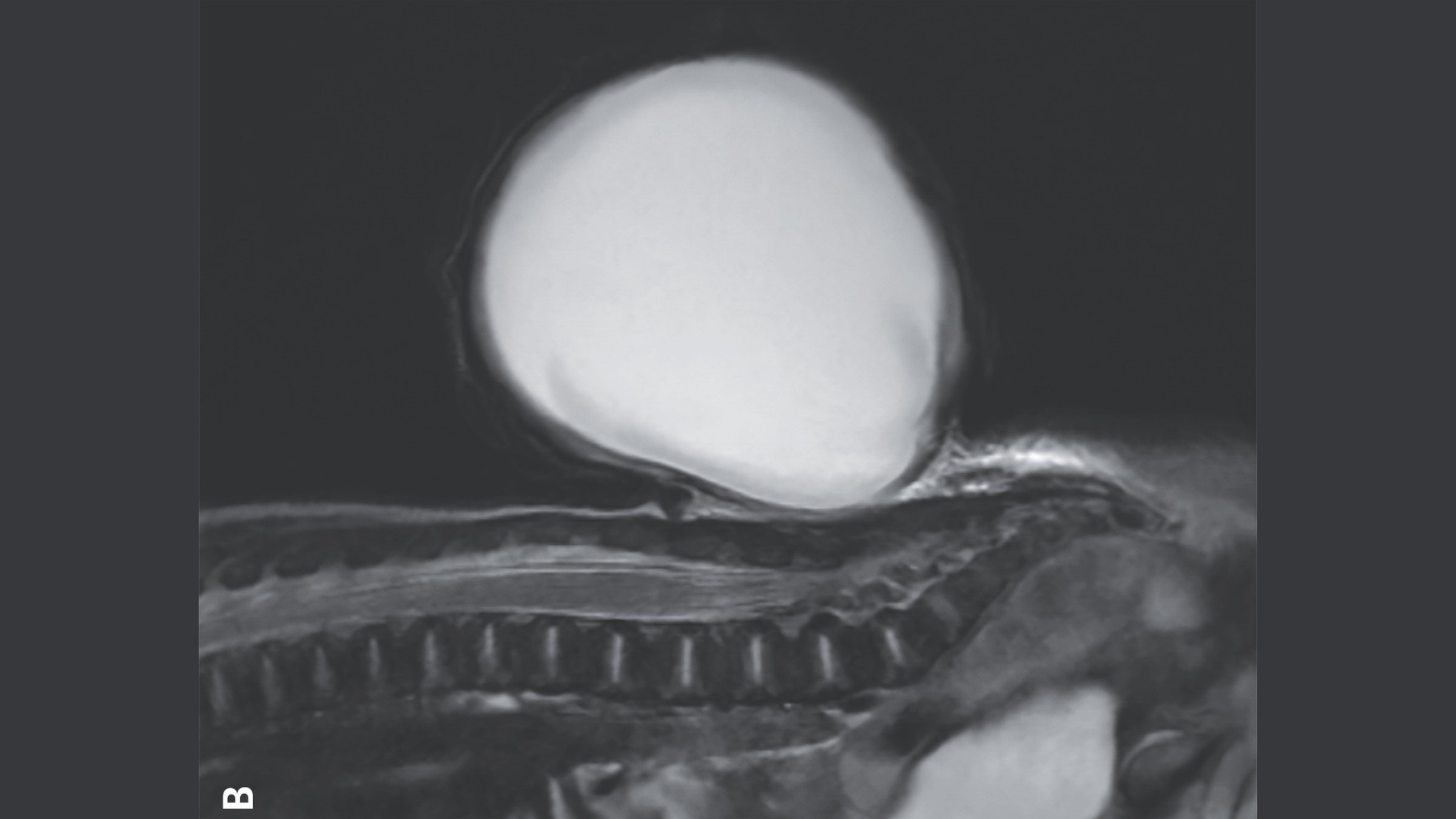
The balloon-like sac of tissue and fluid that grew from the baby’s spine shown on a magnetic resonance imaging (MRI) scan.
The neural metro is a hollow body structure that formsduring the third and quaternary week post - conceptionand it later becomes thebrain and spinal corduroy . Sometimes this process is cut off , depart babe with a col in their spine known asspina bifida . unremarkably , this disruption is covered by skin and does n’t cause any symptoms , and many people areunaware they have the condition .
on occasion , though , tissue paper and fluid that encompass and protects the spinal electric cord is pushed through the gap , create a sac - like , bulge social organisation . This is what happened to the son in the image , who had a specific version of spina bifida called meningocele .
relate : female parent rejoices after her tiddler ’s successful spina bifida surgery in the womb

(Image credit: Live Science)
Scientistsdon’t experience what make spina bifida ; however acombination of genetic , nutritional and environmental risk of infection factorsmake it more probable . For instance , if the female parent does not have enough vitamin M or vitamin B9 during former pregnancy , takes certain medicinal drug such as theanti - epileptic drug valproic acid , or hasdiabetesthat is not well oversee , there is a expectant risk of them having a baby with spina bifida .
However , none of these constituent played a role in the infant ’s condition in this typesetter’s case , harmonise to a report of his eccentric published Dec. 28 , 2024 in theNew England Journal of Medicine .
Doctors first noticed the spinal mar during an ultrasound test around 20 weeks , orhalfway through , the maternity . patient role with meningocele often have pocket-size problems such asissues with their bladder and bowels . However , the condition can usually be cover witha simple operative fixture , eitherbeforeorafter giving birth .
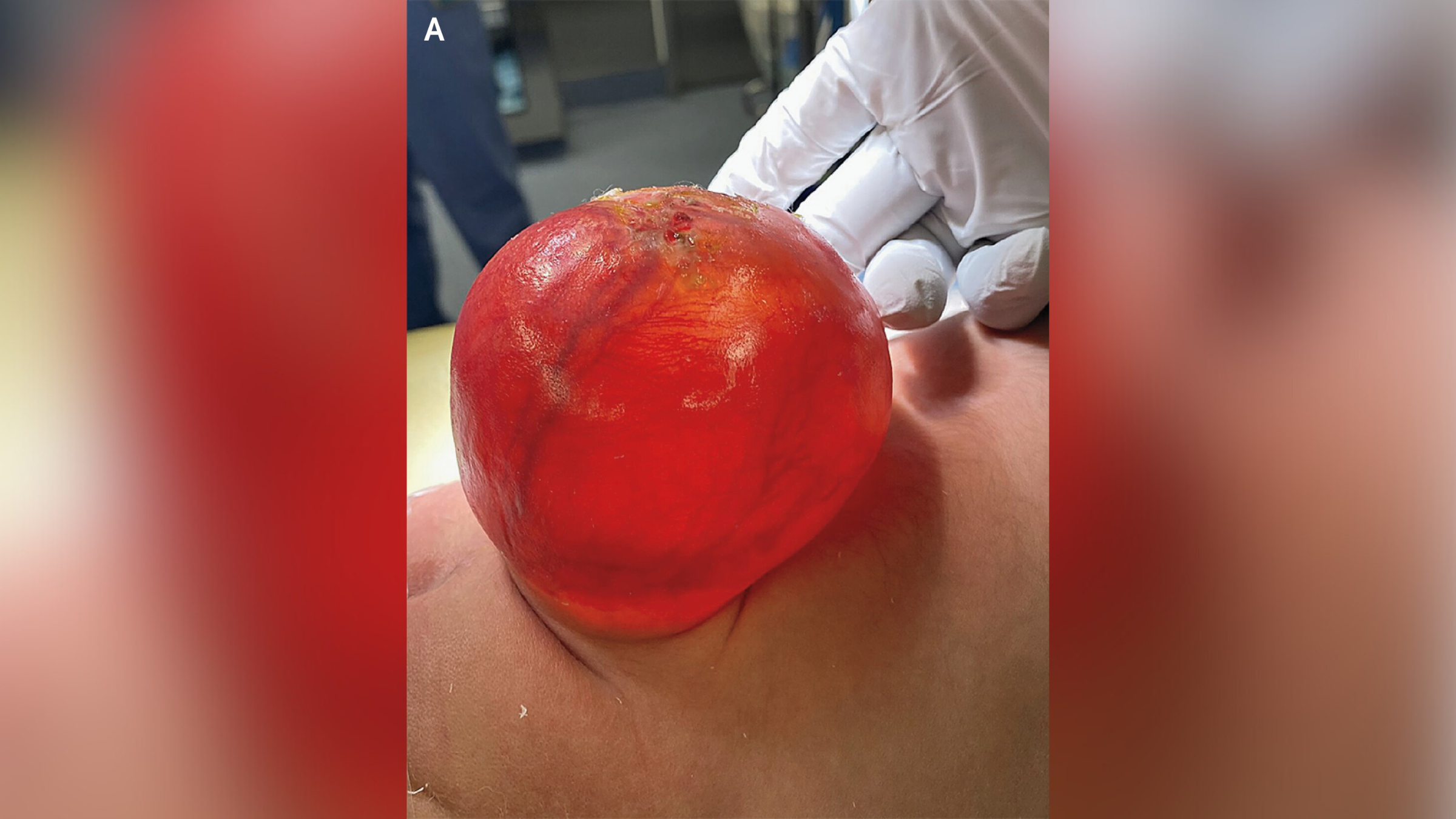
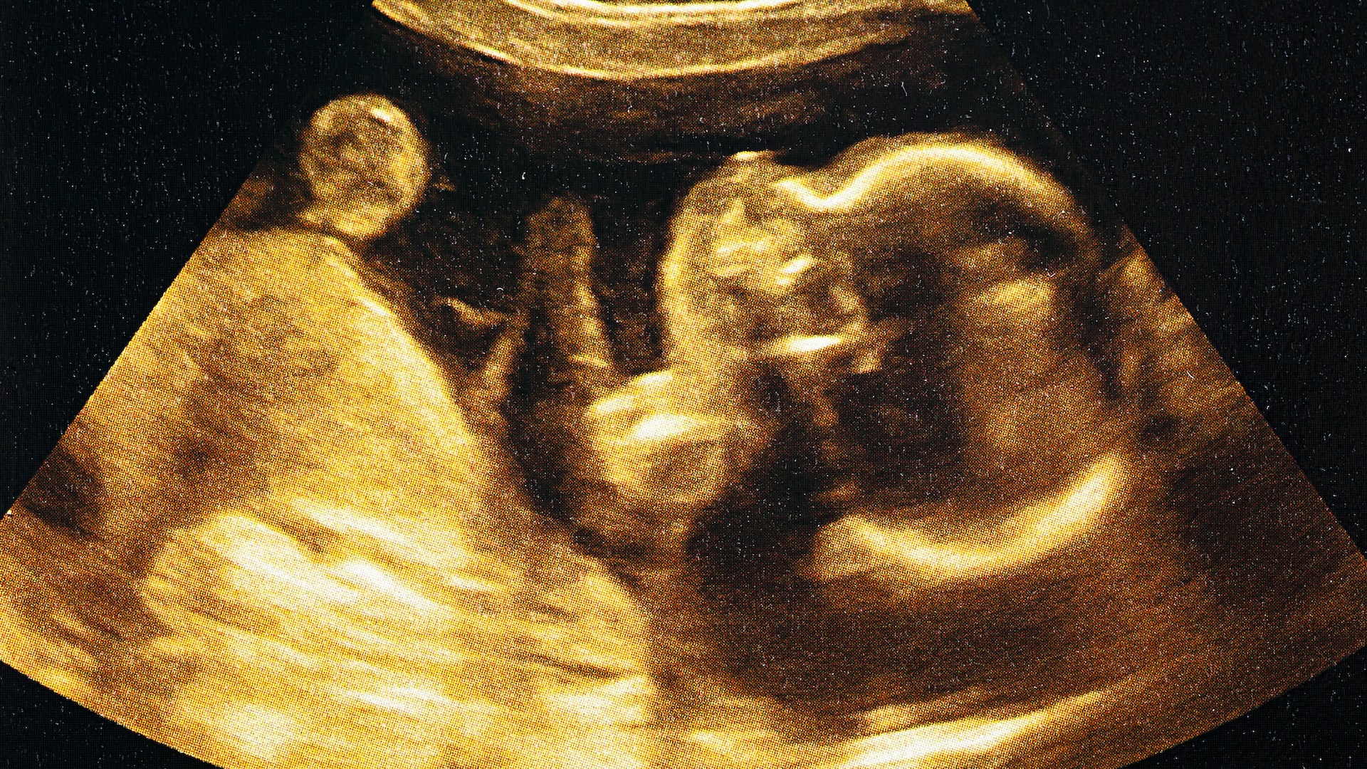
The sac-like protrusion (pictured above) was caused by a condition known as meningocele.(Image credit: The New England Journal of Medicine ©2024)
— In highly rare case , doc remove fetus from brain of 1 - yr - sometime
— Mini model of human embryologic brain and spinal cord grown in lab
— New syndrome identified in children expose to fentanyl in the uterus
Serious complications are more likely to fall out if a babe has another character of spinal bifida make out asmyelomeningocele , in which neural tissue is also establish in the Sauk . For exemplar , these children may be atrisk of arise palsy , frequent urinary nerve tract transmission ( UTIs ) and a type of brain infection known asmeningitis .
In the male child ’s case , his parent opted for him to have surgery to remove the theca and reconstruct his spinal cord after birth . Four 24-hour interval after the surgery , he was discharged home plate , and at his six - month checkup , medico articulate he was developing without any adverse effects .