When you purchase through link on our site , we may make an affiliate committal . Here ’s how it works .
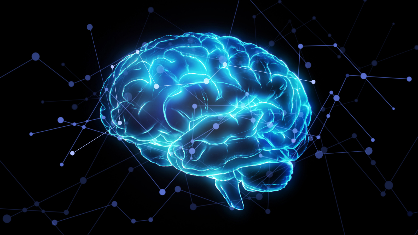
Tiny , hairlike " antennae " protrude from the Earth’s surface of brain cells , and now , scientist have unveil a elaborate map of these wires across the whole human cortex . They go for the new function will guide next inquiry into a year of diseases that stimulate these social organization to malfunction .
The capillary structures , known as cilia , are really found on the control surface of mosteukaryotic cells , imply complex electric cell that house their desoxyribonucleic acid in a karyon . Some cilia can move ; for example , cilia in thelungscollectively beat toclear the airways of harmful pathogens . Others , hollo primary cilia , are immobile and or else act like antennae , sense signals from their environmentand passing them to the nucleus of the cellphone .
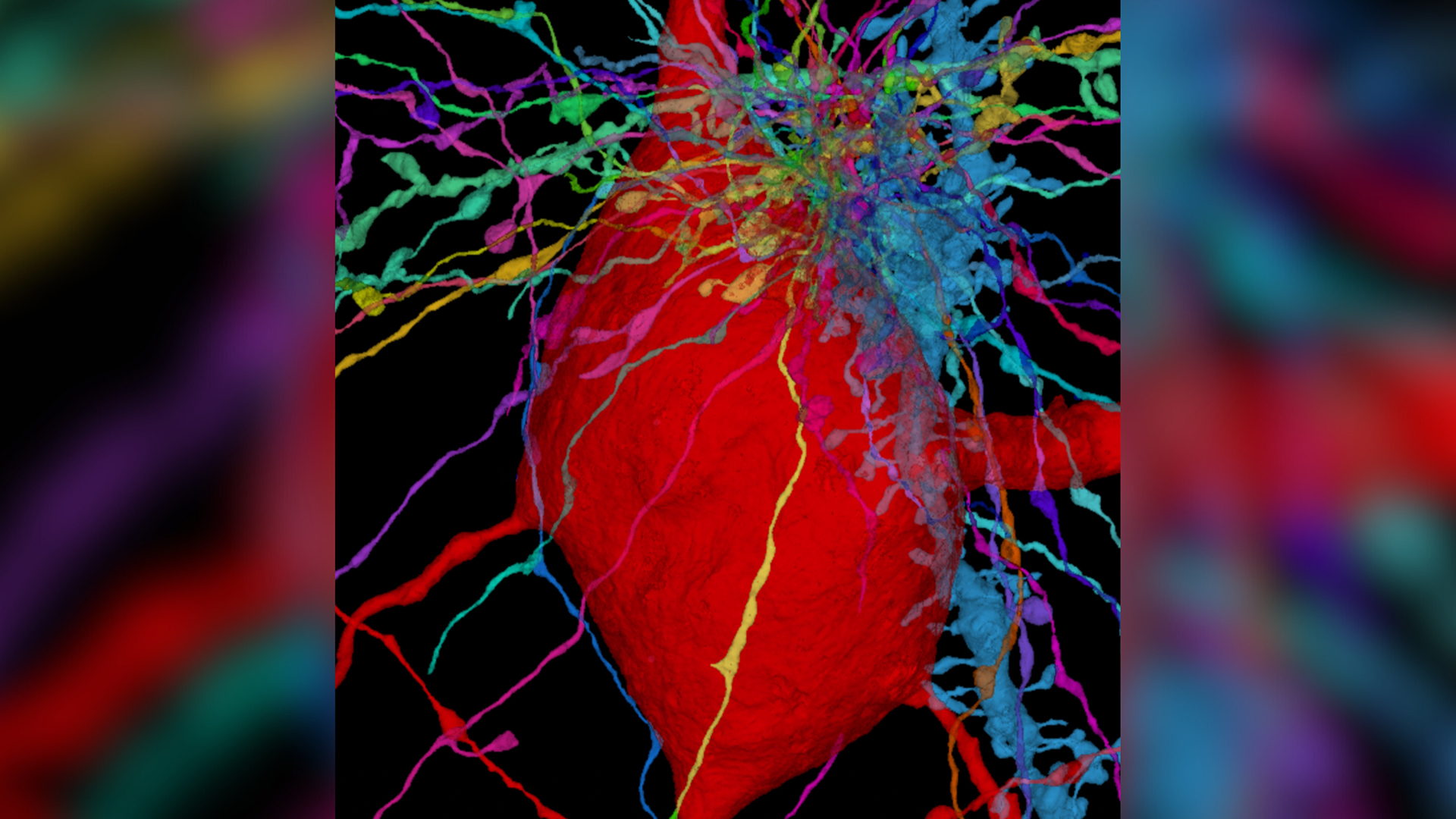
This microscope image shows the “wires” extending from neurons (multicolor) interacting with a specialized type of neuron called an interneuron (red) in the human cerebral cortex. The interneuron has a hairlike antenna called the primary cilium (also red) that’s hidden within the meshwork in the upper-right corner.
In thebrain , nerve cell and their associated supporter cells , calledglia , require primary cilia to effectively receive and send sign . However , until now , little has been known about cilia ’s structure or organization in the brain or how they slot into thenetwork of interactions between neurons in the brainknown as the " connectome . "
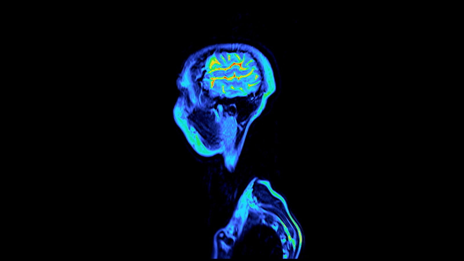
In a new study , published Oct. 27 in the journalNeuron , scientists constructed a three - dimensional single-valued function of the primary cilia in the brain ’s outer level , or cortex , which is part of the cerebrum , the large component of the brain . The researchers hope the new map , which details 56,000 cellular phone , will guide inquiry intociliopathies , a class of disease tied to disruptions in cilia role .
Related : Most elaborated human brain map ever contains 3,300 cell types

" There are a number of cognitive trouble relate with mutations in the proteins that make up lash , " co - senior field of study author , Dr. Jeff Lichtman , a professor of molecular and cellular biology at Harvard University , told Live Science in an e-mail . " These study may suggest that who a cilia contacts may be important in their normal function and when that role goes awry in disease . "
To produce the fresh map , the researchers piece together images of the six layer of the human cerebral cortex ; the prototype depicted " cut " of a tissue sample that had been collected during a surgery to treat an grownup patient role with epilepsy . One by one , the team looked at the cilia that emanated from each mentality prison cell and analyze how they interacted with one another .
The cilia differ in size and shape bet on the type of cell they interacted with and the layer of the cortex where they were place . Cilia were also a key component of the structure of some synapsis — the junctions between nerve cell that allows these cellular telephone tocommunicate with each other — suggesting that cilia are firmly embedded in the brain ’s connectome .

The diversity of the cilia raises the " intriguing possibility " that dissimilar types of cilia in the cortex can order the activity of neurons in unparalleled way , the authors said .
— Newfound ' wit touch ' connect to multiple psychiatric disorders
— Your aboriginal language may form the wiring of your brain

— 1st complete mapping of an insect ’s psyche control 3,016 neurons
go forward , the squad would care to delve further into how these primary cilia influence neural circuits in the brain and how this noesis could be used to treat neurological disorders where cilial function is hinder , co - senior report authorE. S. Anton , a professor of neuroscience at the University of North Carolina School of Medicine , evidence Live Science in an email .
Ever marvel whysome people build musculus more easy than othersorwhy freckle come out in the sun ? send out us your query about how the human body works tocommunity@livescience.comwith the open job " Health Desk Q , " and you may see your question answer on the website !